Loculated Pleural Effusion / Pleural Effusion Amboss. A pleural effusion is due to the manifestations of another illness. A pleural effusion representsthe disruption of the normal mechanisms of formationand drainage of fluid from the pleural space. In vitro efficacy of varidase versus streptokinase or urokinase for liquefying thick purulent exudative material from loculated empyema. Loculated pleural effusion the pleura is a thin membrane between the lungs and chest wall that lubricates these surfaces and allows movement of the lungs while breathing. Lung scarring and a permanent decrease in lung function are associated with chronic pleural effusion.
Loculated malignant effusions however, are inherently resistant to the usual approaches because of nonexpanding underlying lung. If it is clear that there are multiple loculations then it is wise to avoid delay and proceed directly to this procedure. The lack of specificity is mainly due to the limitations of the imaging modality. Diffuse nodules and opacification in right lung with compressive atelectasis. Twenty of the 21 complicated parapneumonic effusions (including empyemas) showed loculation (95%).
Evaluation Of The Patient With Pleural Effusion Cmaj from www.cmaj.ca Fibrotic scar tissue may form in the pleural cavity (called loculation), preventing effective drainage of the fluid. In vitro efficacy of varidase versus streptokinase or urokinase for liquefying thick purulent exudative material from loculated empyema. This type of effusion is empyema unless proven otherwise. Most malignant effusions can be controlled by thoracentesis and/or closed thoracostomy tube drainage and sclerosis of the pleural cavity. Nonmalignant pleural effusions (nmpes) have a wide variety of etiologies ( table 1 and table 2 and table 3) and cause significant morbidity and mortality 2,3 . Left pleural effusion with high density material at the posterior costophrenic angle. Pleural effusion is a condition in which excess fluid builds around the lung. Treatment may fail if the catheter is not placed optimally within the loculation or if the fluid is hemorrhagic or fibrinous.
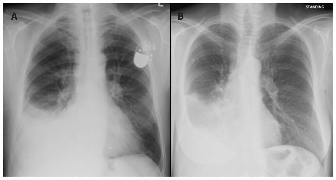
The largest pocket of fluid is present posteriorly at the right lung base, with associated atelectasis and minor consolidation.
Icu patients cannot sit up and the effusion layers posteriorly. Left pleural effusion with high density material at the posterior costophrenic angle. Most malignant effusions can be controlled by thoracentesis and/or closed thoracostomy tube drainage and sclerosis of the pleural cavity. A pleural effusion occurs when fluid fills this gap and separates the lungs from the chest wall. Pleural effusions describe fluid between the two layer of tissue (pleura) that cover the lung and the lining of the chest wall. Treatment may fail if the catheter is not placed optimally within the loculation or if the fluid is hemorrhagic or fibrinous. In chf effusions are bilateral and more on right. Lung scarring and a permanent decrease in lung function are associated with chronic pleural effusion. Pleural effusion that is confined to one or more fixed pockets in the pleural space. Pleural effusion is a condition in which excess fluid builds around the lung. The pleura are thin membranes that line the lungs and the inside of the chest cavity and act to lubricate and facilitate breathing. Pleural fluid is seen extending to the right oblique fissure. Normally, a small amount of fluid is present in the pleura.
A rationaldiagnostic workup, emphasizing the most commoncauses, will reveal the etiology in most cases. A pleural effusion representsthe disruption of the normal mechanisms of formationand drainage of fluid from the pleural space. The pleura are thin membranes that line the lungs and the inside of the chest cavity and act to lubricate and facilitate breathing. The largest pocket of fluid is present posteriorly at the right lung base, with associated atelectasis and minor consolidation. Twenty of the 21 complicated parapneumonic effusions (including empyemas) showed loculation (95%).
Tuberculous Pleural Effusion Brown Emergency Medicine from images.squarespace-cdn.com Pleural effusion that is confined to one or more fixed pockets in the pleural space. Pleural effusion is when fluid fills this gap and separates the lungs from the chest wall. Loculated pleural effusion the pleura is a thin membrane between the lungs and chest wall that lubricates these surfaces and allows movement of the lungs while breathing. What are the different appearances of pleural effusion? Surgical thoracostomy tube placement and radiologically guided catheter drainage are standard therapy for loculated pleural fluid collections. Nonmalignant pleural effusions (nmpes) have a wide variety of etiologies ( table 1 and table 2 and table 3) and cause significant morbidity and mortality 2,3 . Icu patients cannot sit up and the effusion layers posteriorly. Twenty of the 21 complicated parapneumonic effusions (including empyemas) showed loculation (95%).
Surgical thoracostomy tube placement and radiologically guided catheter drainage are standard therapy for loculated pleural fluid collections.
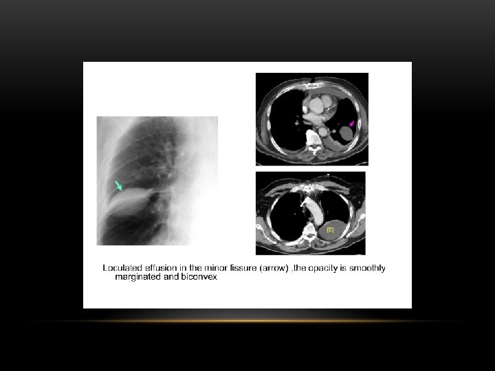
Indwelling pleural catheters (ipcs) are effective management options for malignant pleural effusion. Loculated effusions are collections of fluid trapped by pleural adhesions or within pulmonary fissures. Left pleural effusion with high density material at the posterior costophrenic angle. The pleura are thin membranes that line the lungs and the inside of the chest cavity and act to lubricate and facilitate breathing. Pleural effusions describe fluid between the two layer of tissue (pleura) that cover the lung and the lining of the chest wall. Pleural fluid is seen extending to the right oblique fissure. Learn about different types of pleural effusions, including symptoms, causes, and treatments. There are no established guidelines to facilitate management of nmpes and most management strategies rely on expert experience and data derived from patients with malignancy. Pleural effusion that is confined to one or more fixed pockets in the pleural space. What are the different appearances of pleural effusion? Fibrotic scar tissue may form in the pleural cavity (called loculation), preventing effective drainage of the fluid. This type of effusion is empyema unless proven otherwise. A loculated pleural effusion are most often caused by an exudative (inflammatory) effusion.
This more efficient drainage improved dyspnoea more rapidly, but. Surgical thoracostomy tube placement and radiologically guided catheter drainage are standard therapy for loculated pleural fluid collections. In chf effusions are bilateral and more on right. Pleural fluid is seen extending to the right oblique fissure. There are no established guidelines to facilitate management of nmpes and most management strategies rely on expert experience and data derived from patients with malignancy.

0 Response to "Loculated Pleural Effusion / Pleural Effusion Amboss"
Post a Comment